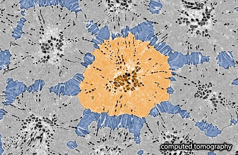
How are tesserae interacting with one another?
We learned from our developmental study that tesserae grow and become in contact in sub-adult stages. From this moment on, intertesseral joints form complex architectures, comprised of regions of intimate abutment of the two opposing edges of adjacent tesserae (intertesseral contact zones, icz) and small gaps containing cells and fiber bundles (intertesseral fiber zones (ifz).
The combination of mineralized tissue and unmineralized intertesseral joints is vital to elasmobranch skeletal biology, providing both cortical stiffness for mechanical function and room for interstitial growth between tesserae. We presented a quantitative study of the intertesseral joint architecture at SICB 2018 (Seidel et al. 2018. Quantitative shape analysis and mechanics of intertesseral joints in tessellated cartilage of sharks and rays. Integrative and comparative Biology. Vol. 58. Journals Dept. 2001 Evans Rd, Cary, NC 27513 USA: Oxford University Press Inc, 2018).
Tesserae appear not to fuse, suggesting that some mineralization inhibition of the fibrous tissue between adjacent tesserae exists, which is likely orchestrated by the cells in these regions. The growth and mechanics of tessellated cartilage skeletons rely on the maintenance of linked, but separated, tiles, and in Seidel et al. 2016 we provide illustrations and discussed the processes involved in joint development and maintenance during skeletal growth. (Seidel et al., 2016. Ultrastructural and developmental features of the tessellated endoskeleton of elasmobranchs (sharks and rays). Journal of Anatomy 229.5: 681-702)


