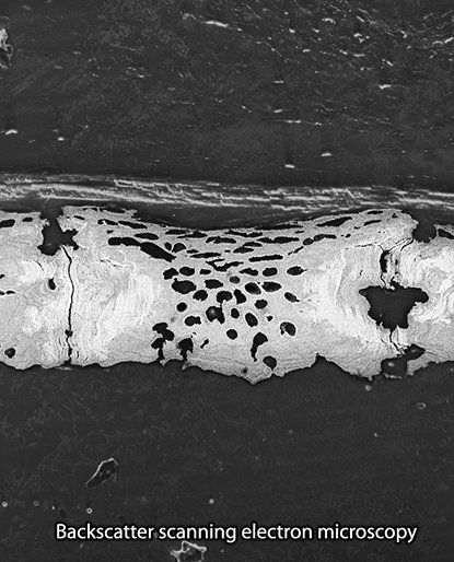
Ultrastructural features of tesserae
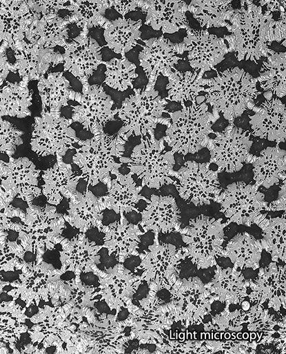
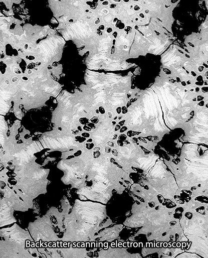
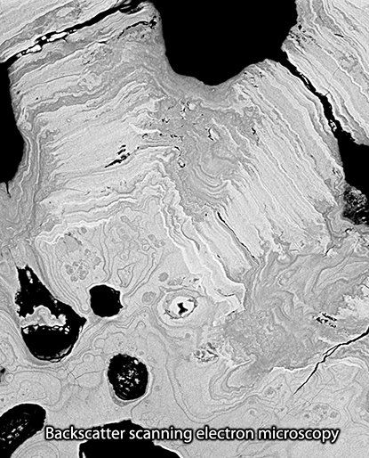
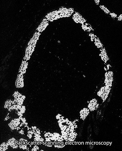
Tesserae are more than just blocks of mineral! Across species, but also within skeletal elements of the same indiviual tesserae exhibit divers shapes. Our ultrastructural study of tesserae, in particular of those from round stingray Urobatis halleri, revealed several important features of tesseral anatomy. For example tesseral spokes, lamellated, high-mineral density features radiating outward from the center of each tessera to its neighbors, like spokes on a wheel, likely acting as structural reinforcements of the articulations between tesserae.
This and other anatomical features were described in Seidel et al. 2016 and are common among species of all major elasmobranch groups despite the large variation in tesseral shape and size. Most of them relate to local variations in grey values in backscatter images, which are variations in mineral density. See some backscatter electron images below or have a look at our publication (Seidel et al., 2016. Ultrastructural and developmental features of the tessellated endoskeleton of elasmobranchs (sharks and rays). Journal of Anatomy 229.5: 681-702.) where we discuss hypotheses about how these features develop, and compare them with other vertebrate skeletal tissue types and their growth mechanisms.


